Micro-Image Your Research
Student photomicrography celebrated in “Micro-Image Your Research” event
The Electron Microscopy Lab, in conjunction with the Innovative Media Research and Commercialization Center, both members of the University of Maine’s Coordinated Operating Research Entities (CORE), invited student researchers to consider a new perspective on their specimens this semester.
“Micro-Image Your Research” invited undergraduate and graduate students from a variety of areas of study to select a specimen and use photomicrography to capture unique images at high levels of magnification that would help to communicate new understandings about their research. Student contestants represented disciplines within the School of Marine Science, School of Earth and Climate Sciences, School of Biology and Ecology, the Ecology and Environmental Sciences Program, and the Departments of Anthropology and History.
For many students and their advisors, it was their first use of the Electron Microscopy Lab. “The competitors were from labs or departments that do not usually work with the Electron Microscopy CORE and did not know much about it. The competition brought the facility to their attention and showed them how applicable it could be for their research,” noted laboratory manager Emma Perry. “Students who entered were very excited about their experience.”
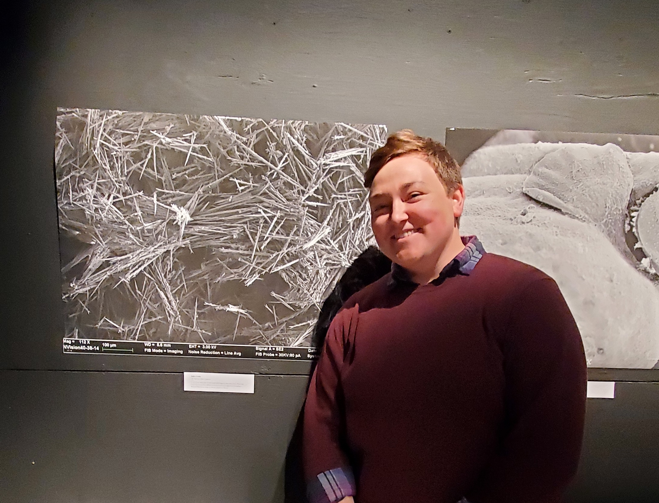
The contest culminated in a gallery reception at the IMRC Center on the evening of April 24th. Each image was printed in large format courtesy of the IMRC Center’s Prototyping Lab to further emphasize magnified details. Four guest judges, all current or retired UMaine faculty and staff, rated each entry on composition, originality, and impact: Sheridan Adams, Director of Instructional Design & Support Services for the Center for Innovation in Teaching and Learning (CITL) and Adjunct Assistant Professor of the Intermedia MFA Program; Bill Küykendall, retired Senior Lecturer in New Media and Cooperating Professor of Communication and Journalism; Roger Merchant, forester and retired Cooperative Extension educator; and Patrick Wine, Division of Marketing and Communications Photographer and Videographer.
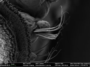
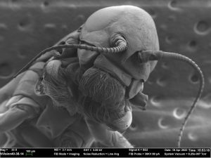
The top three contestants were announced at the event: in 3rd place, Emma Thomasetti, School of Marine Sciences, advised by Paul Rawson, with images of the Polydora websteri polychaete worm; in 2nd place, Val Watson, School of Biology and Ecology, advised by Hamish Grieg, with images of the Isonychia mayfly nymph and Leptoceridae caddisfly larva; and in 1st place, Sadia Crosby, School of Biology and Ecology, advised by Angela Mech, with images of Euproctis chrysorrhoea Browntail Moth hairs.
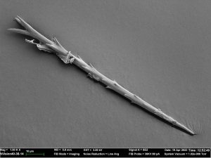
The contest served to highlight the importance of inspiring new understandings where creative activity and research meet. Of Crosby’s winning images, a judge stated: “I immediately get the collaborative context, visual and words, conveyed by these two photos”; “The lighting is stunning and provides an ominous look at these little spines,” another judge wrote. “I would gladly hang this image in a gallery or home.”
UMaine CORE thanks all contestants, advisors, judges, and EML and IMRC Center staff for their coordination on this effort. The contest is anticipated to return next year, and will continue to be open to both graduate and undergraduate students. CORE resources are available to the entire UMaine community; learn more at umaine.edu/core.